Diagram Of Animal Cell Under Electron Microscope Labeled. Label the cell wall cell-surface membrane capsule circular DNA flagella and plasmid.
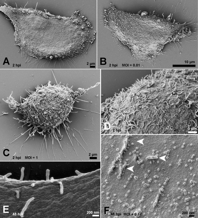
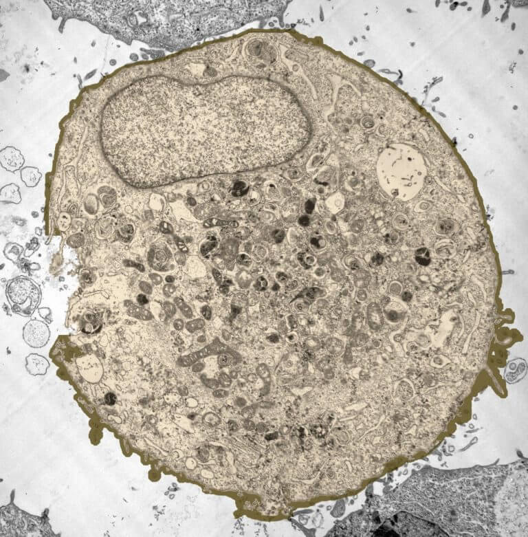
Ultrastructural Analysis Of Sars Cov 2 Interactions With The Host Cell Via High Resolution Scanning Electron Microscopy Scientific Reports
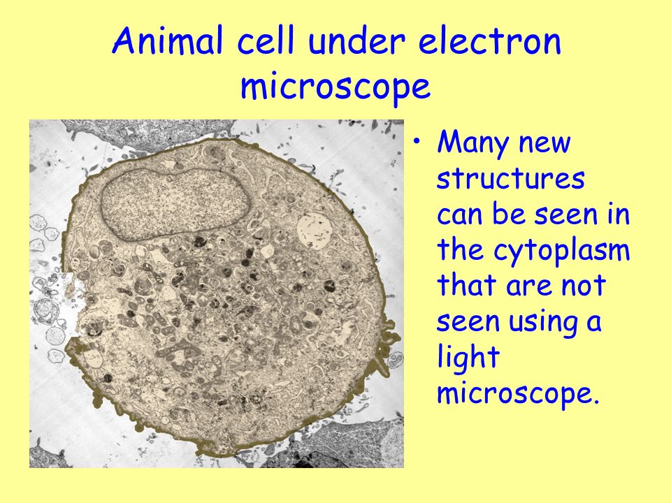
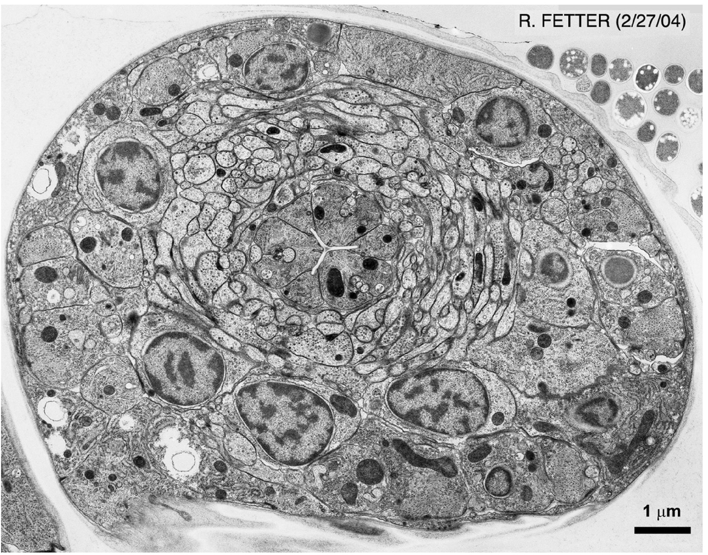
Under the intense radiation of the electron microscope 011 electron per Å 2 the question of viability of cells naturally arises because the amount of radiation absorbed during highmagnification imaging is sufficient to cause cell death.
Animal cell under electron microscope labelled. The cell membrane is important in that. Labeled animal cell under electron microscope 8745961 orig. Animal cells have a basic structure.
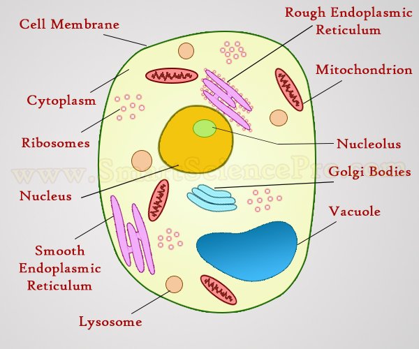
Make your work easier by using a label. Describe why it is important to prepare. Endoplasmic Reticulum Rough And Smooth British Society For.
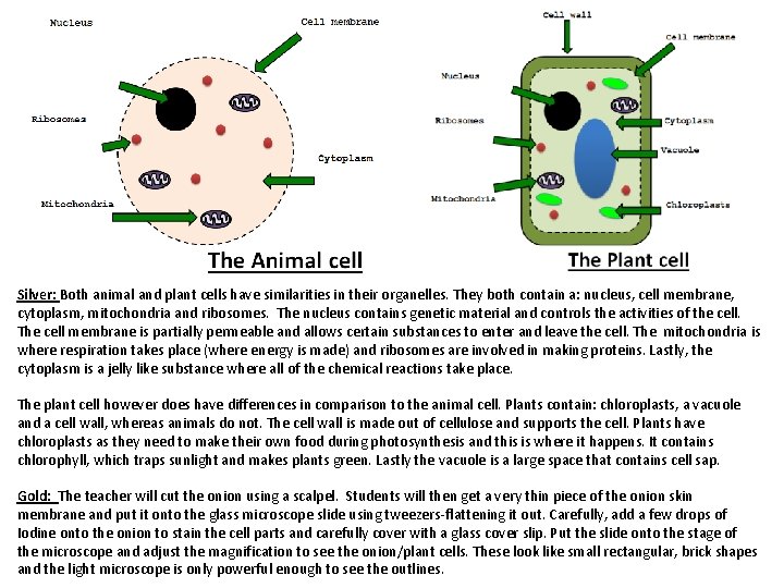
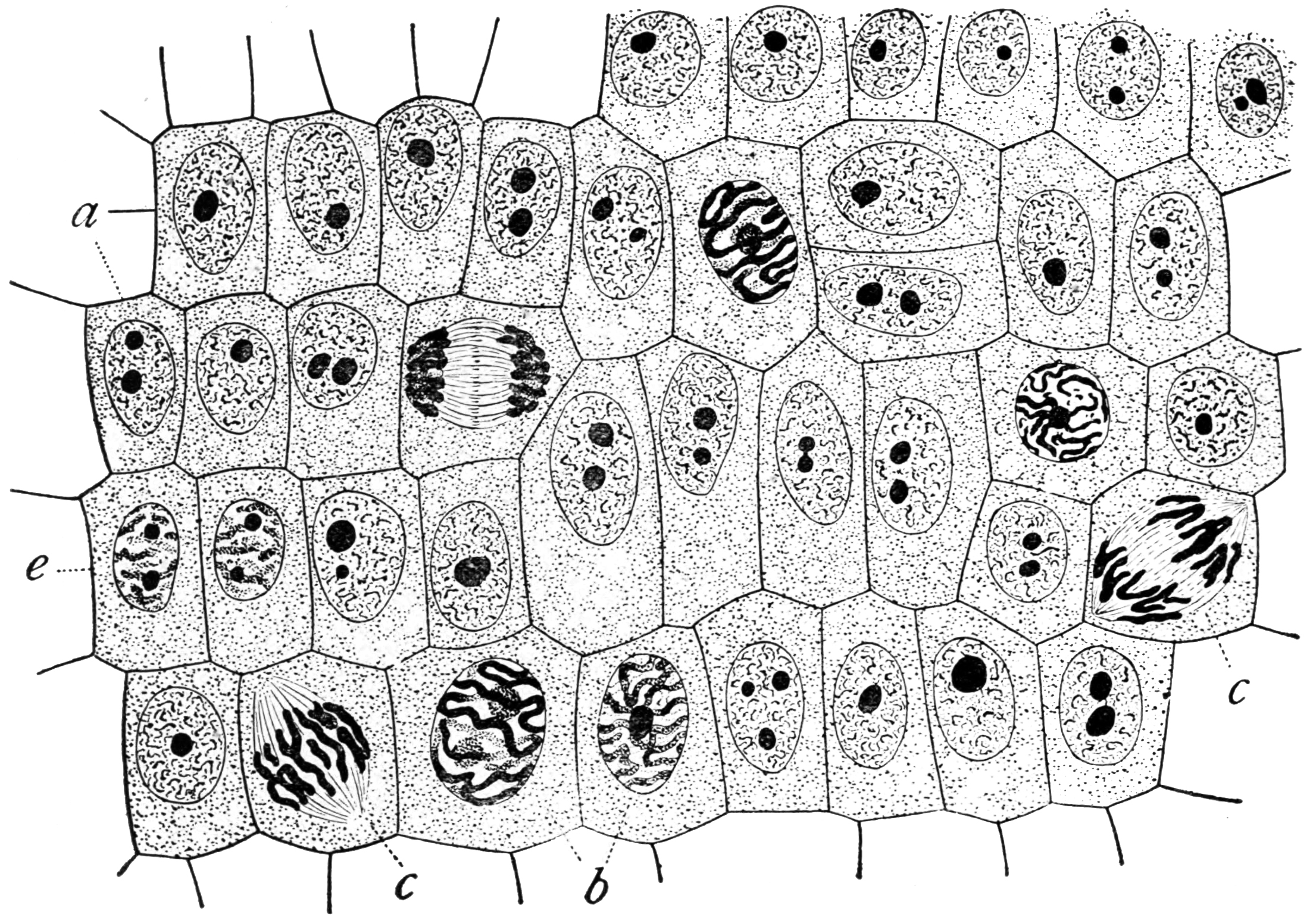
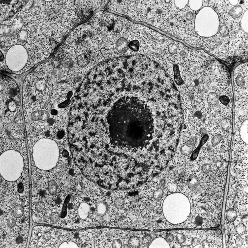
Labelled diagram of a plant cell under microscope posted on march 18 2011 by admin onion cells stained with methylene blue look at the images of onion cells as they would be seen under a microscope draw each magnification label appear high picture plant and animal cell. But at the same time it is interpretive. Labeled Animal Cell Under Electron Microscope.
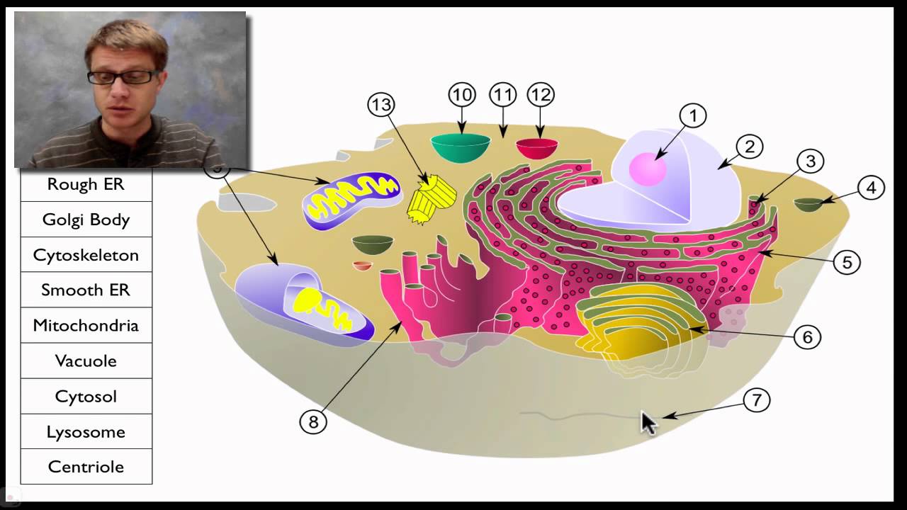
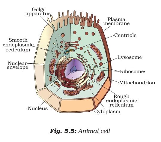
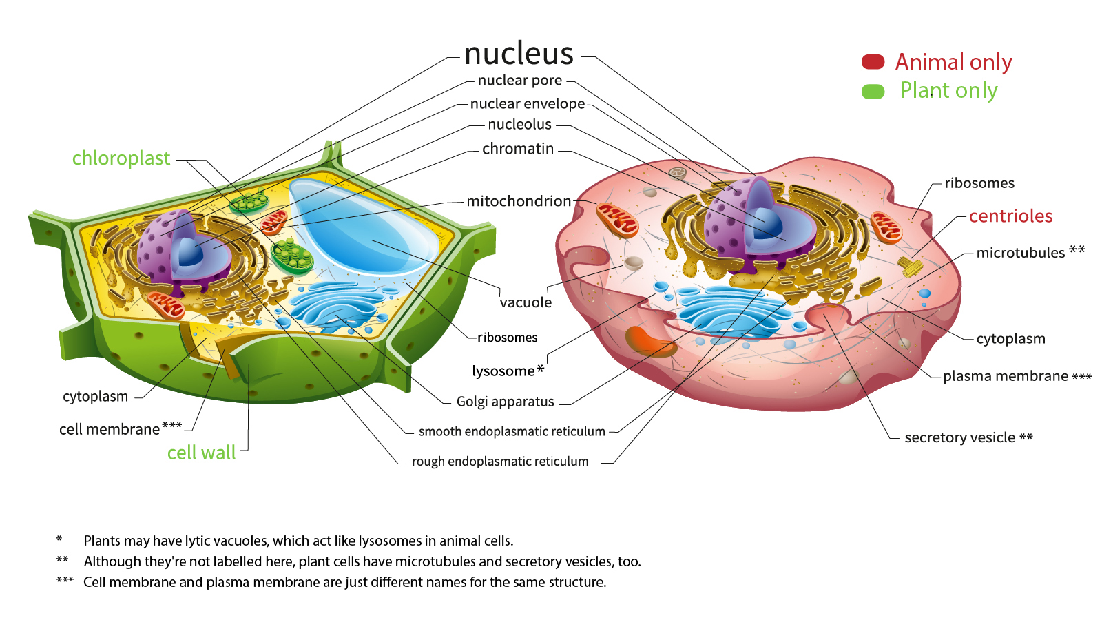
Draw a labelled diagram of the internal structure of an animal cell as seen with an electron microscope. Animal Plant Cells Gcse Science Biology Get To Know Science Youtube Mitochondrion are visible with a light microscope but cant be seen in detail. Ribosomes are only visible with an electron.
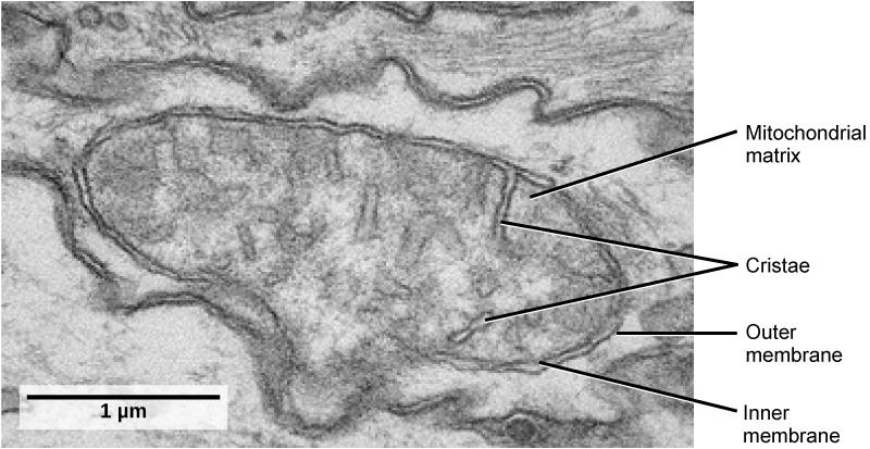
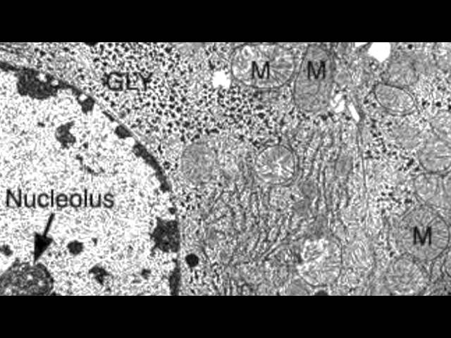
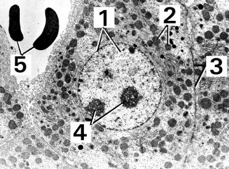
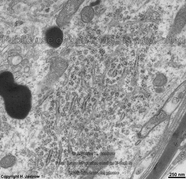
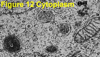
The original and the labelled images are already used world-wide for preparation for exams. Labels are a means of identifying a product or container through a piece of fabric paper metal or plastic film onto which information about them is printed. It is an electron micrograph of cells largest and most important organelle the mitochondria and is characterized by the following features Fig.
Bookfanatic89 Diagram Of Plant Cell Under Electron Microscope. Make your work easier by using a label. Its a thin slice.
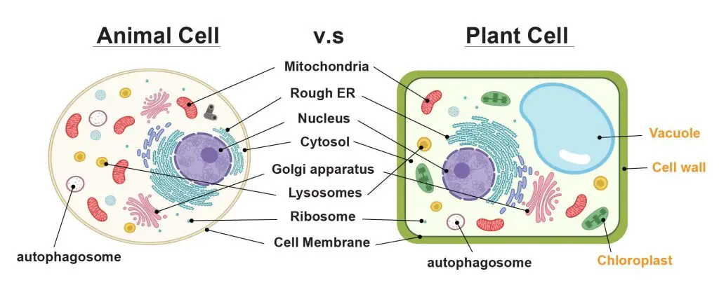
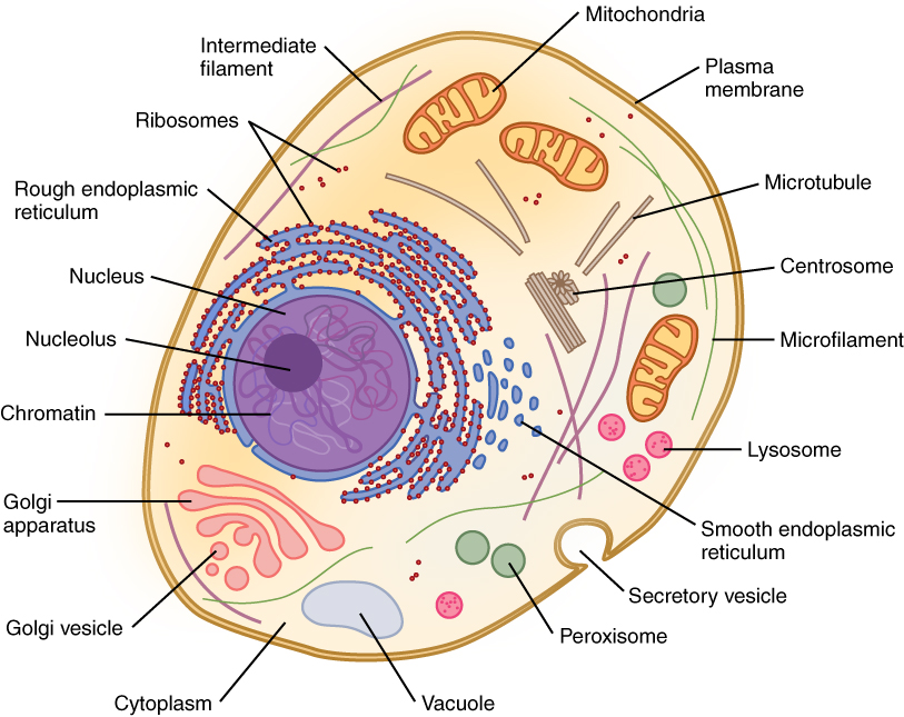
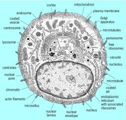
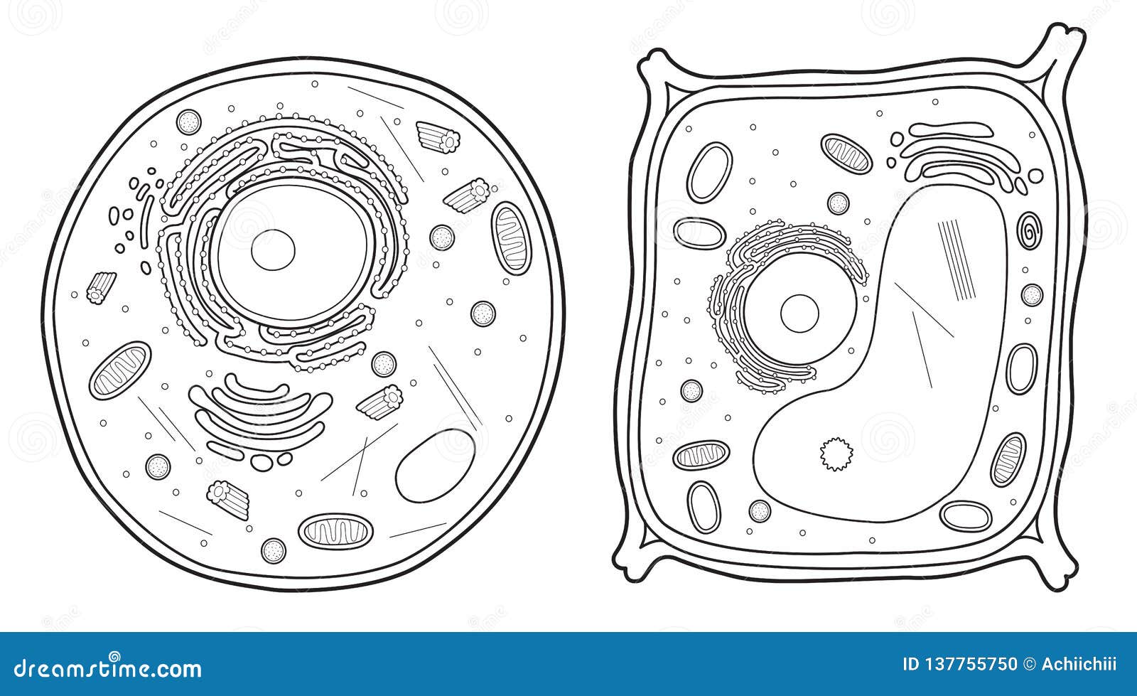
Clearly visualized under an electron microscope it must be labeledcell structure the physics teacher april 23rd 2018 - 2 1 cell structure identify the parts of an animal cell as seen under light microscope the existence and definition of prokaryotic and eukaryotic cells me 5 10. So lets begin by drawing a rough-oval shape. See how a generalized structure of an animal cell and plant cell look with labeled diagrams.
Ziehen die pins an die richtige stelle auf dem bild. It is flexible and has pores. However no obvious structural damage.
We all do not forget that the human physique is quite problematic and a method I. Diagram Of Animal Cell Under Electron Microscope. Both the global and high-resolution distribution of colloidal gold labels on cells can be readily determined.
Labels are a means of identifying a product or container through a piece of fabric paper metal or plastic film onto which information about them is. The plant cell as more rigid and stiff walls. Posted 6 years ago.
Wide collections of all kinds of labels pictures online. Wide collections of all kinds of labels pictures online. These are both specific types of.
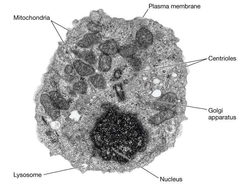
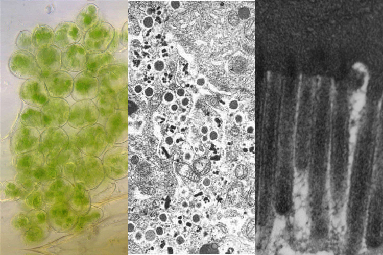
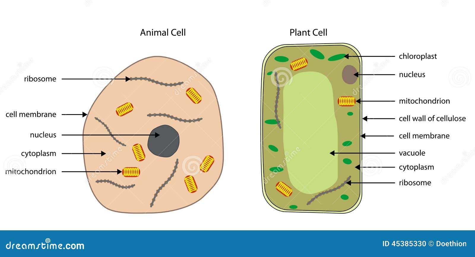
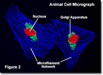
Learn the structure of animal cell and plant cell under light microscope. You see that many features are in common. Here is an electron micrograph of an animal cell with the labels superimposed.
Labelled animal cell diagram gcse. The cell membrane also known as plasma membrane or plasmalemma consists of three layers when viewed under the electron microscope. Electron Micrograph Animal Cell Under Electron Microscope.
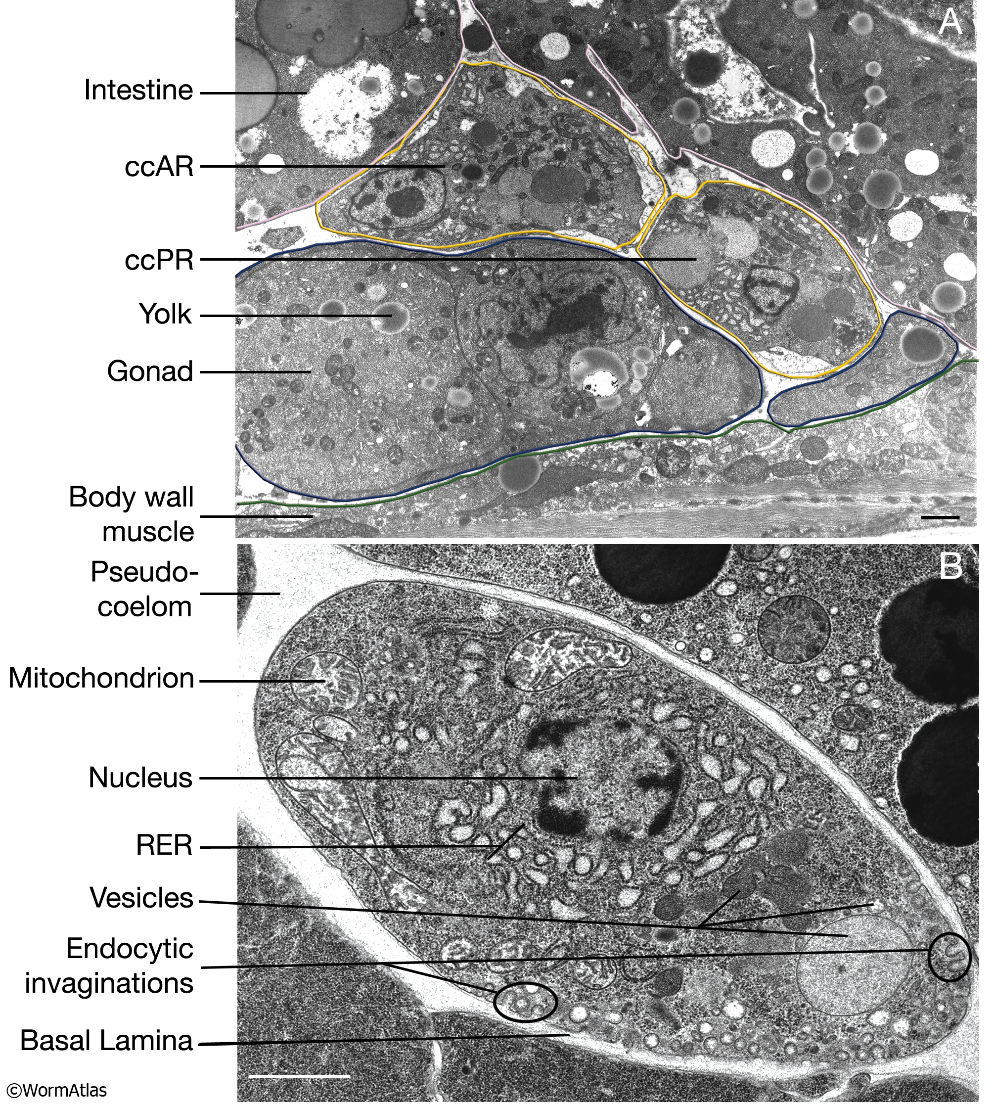
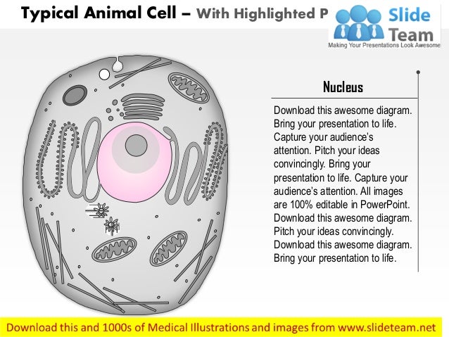
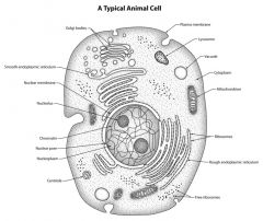
A typical animal cell as seen in an electron microscope Medical Images For PowerPoint. Most cells both animal and plant range in size between 1 and 100 micrometers and are thus visible only with the aid of a microscope. Transmission and some scanning electron microscopic images oforgans cells.
Monday April 5th 2021. The three layers are composed of one layer of phospholipid sandwiched between two protein layers. Table D leads to images of electron microscopes or protocols for tissue preparation.
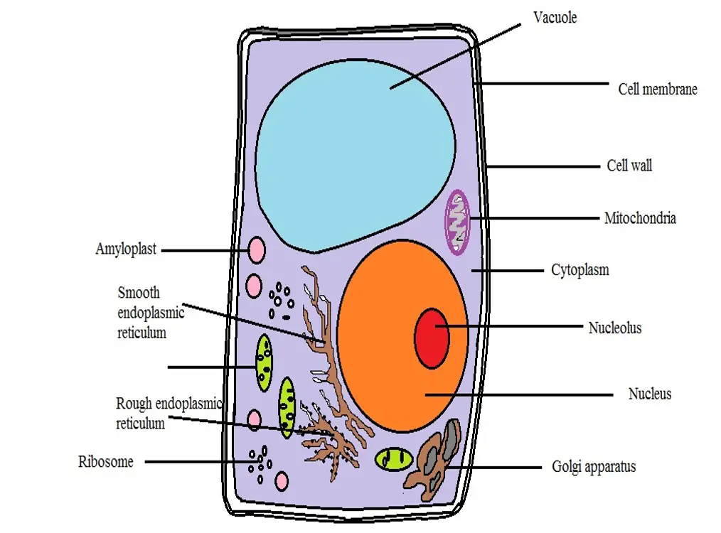
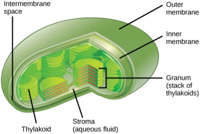
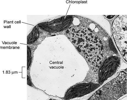
1 The name mitochondria was given by Benda 1898 and their ma n function was brought to light by Kingsbury 1912. Plant Cell Under Electron Microscope Labelled Written By MacPride Tuesday June 18 2019 Add Comment Edit. Draw a labelled diagram of the internal structure of a plant cell as seen with an electron microscope.
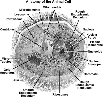
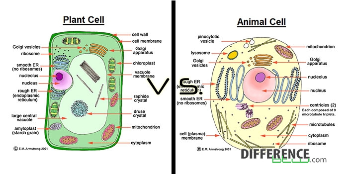
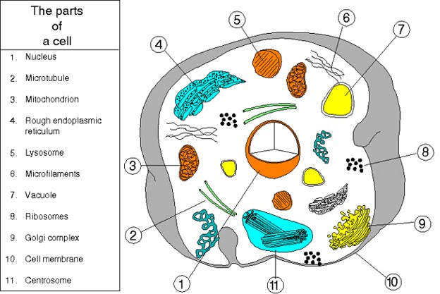
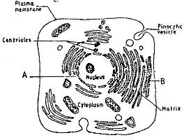
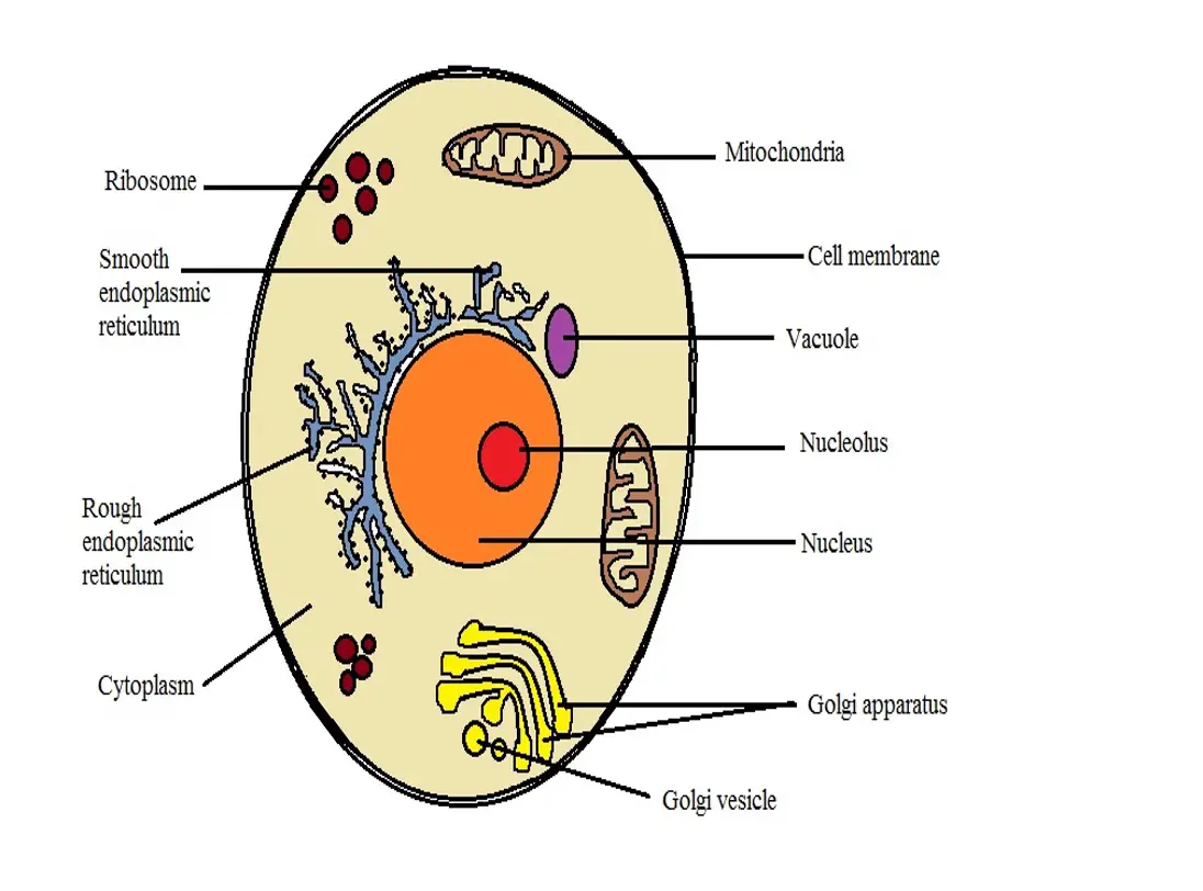
Typical Animal Cell Pinocytotic vesicle Lysosome Golgi vesicles Golgi vesicles rough ER endoplasmic reticulum Smooth ER no ribosomes Cell plasma membrane Mitochondrion Golgi apparatus Nucleolus Nucleus Centrioles 2 Each composed of 9 microtubule triplets Microtubules. The information can be in the form of hand-written or printed text or. The diagram is very clear and labeled.
The animal cell is more fluid or elastic or malleable in structure. The lack of a rigid cell wall allowed animals to develop a greater diversity of cell types tissues and organs. Below the basic structure is shown in the same animal cell on the left viewed with the light microscope and on the right with the transmission electron.
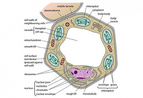
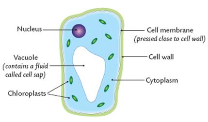
2 Each mitochondria in section appears as sausage or cup or bowl shaped structure lined by double. A brief explanation of the. Heres a diagram of a plant cell.
Cell is a tiny structure and functional unit of a living organism containing various parts known as organelles.

2 3 3 Identify Structures From Electron Micrographs Of Liver Cells Youtube
Draw A Neat Labelled Diagram Of An Animal Cell Studyrankersonline
A Cell Unit Of Function Biology4isc
3 3 Eukaryotic Cells Concepts Of Biology 1st Canadian Edition

Organelles Biology For Majors I
Chapter 7 Introduction To Cells Introduction To Molecular And Cell Biology

Electron Microscope Images Of Rat Liver Cell Changes Control Group Download Scientific Diagram

Plzz Answer This Q Q 19 Given Below Is A Diagrammatic Sketch Of Electron Microscopic View Of An Animal Science The Fundamental Unit Of Life 12392108 Meritnation Com
Cell Types And Organelles

1
Introduction To Cell

Eukaryotic Cells Types And Structure With Diagram

Aice Biology Chapter 1 Plant Cell Electron Micrograph Labeling Diagram Quizlet
![]()
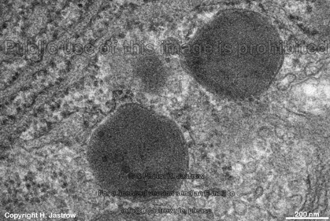
Transmission Electron Micrograph Tem Of Lysosomes A Lysosome Is A Membrane Bound Organelle Found In Nearly All Animal Cells They Are Spherical Vesicles Which Contain Hydrolytic Enzymes That Can Break Down Virtually All

Anatomy And Physiology Of Animals The Cell Wikibooks Open Books For An Open World
Animal Cell Structure Diagram Model Animal Cell Parts And Organelles With Their Functions Jotscroll
Eukaryotic Cells Definition Eukaryotic Cell Diagram Parts Structure And Examples Jotscroll
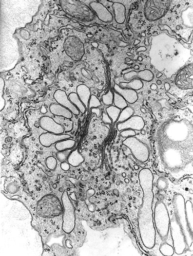
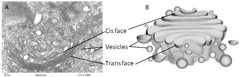
Golgi Apparatus British Society For Cell Biology

Learning Activity 1 1 Learning About Cells And Microscopes
Ht 6 Cells Lesson 1 Microscopy Understand How
Topic 1 2 Ultra Structure Of Cells Amazing World Of Science With Mr Green
Illustrate Only A Plant Cell As Seen Under Electron Microscope How Is It Different From Animal Cell Studyrankersonline
Cell Organelles Science Learning Hub
Cell Structure
Plant Cell Definition Characteristics Facts Britannica
Cell And Organelles Dr Jastrow S Electron Microscopic Atlas
Cell Structure Teaching Resources The Science Teacher
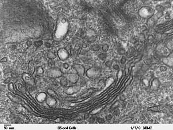
Golgi Apparatus Wikipedia
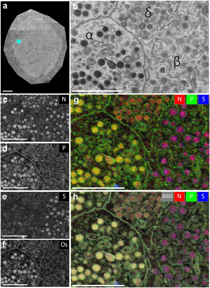
Multi Color Electron Microscopy By Element Guided Identification Of Cells Organelles And Molecules Scientific Reports
1
1
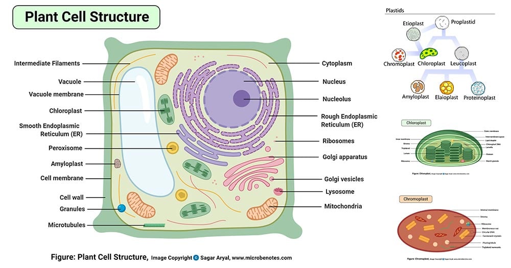
Plant Cell Definition Labeled Diagram Structure Parts Organelles

Cytology Advance Level Notes
Generalized Plant Cell

Ccfig 5 Legend

Electron Microscopes Cell Structure Edexcel Gcse Combined Science Revision Edexcel Bbc Bitesize
4 3 Eukaryotic Cells
Cell Organelles And Their Functions Rs Science

Cell Deck Function By Howard Dunfee
What Is A Diagram Of A Plant And Animal Cell Under An Electron Microscope Quora

Draw The Diagram Of An Animal Cell As Seen Through An Electron Microscope And Label The Parts That Brainly In
Topic 1 2 Ultra Structure Of Cells Amazing World Of Science With Mr Green

Cells Plasma Pancreatic Acinar Ovarian Endocrine Visual Histology
A Typical Animal Cell As Seen In An Electron Microscope Medical Ima
2 3 3 Identify Structures From Electron Micrographs Of Liver Cells Youtube
Cell And Organelles Dr Jastrow S Electron Microscopic Atlas
A Typical Animal Cell As Seen In An Electron Microscope Medical Ima

Cells And Microscopy Diagram Quizlet
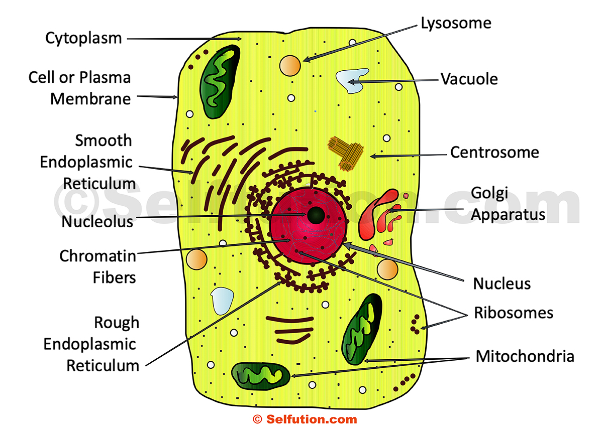
Structure Of Generalized Cell Plant And Animal Selftution

Structure Of Animal Cell And Plant Cell Under Microscope Diagrams Cell Diagram Animal Cell Plant Cell Diagram

Mohammed Awal Mohammedawal950 Profile Pinterest
Cell Biology Wikipedia

Monster Designs Animal Cell Under An Electron Microscope

Non Membranous Organelles And Membranous Organelles In The Cytoplasm Science Online
The Nucleus And Cytoplasm Anatomy And Physiology
Cell Structure Learning Intention Ppt Video Online Download

1 2 Skill Interpretation Of Electron Micrographs Youtube
Difference Between Plant And Animal Cells Cells As The Basic Units Of Life Siyavula
2 3 Eukaryotic Cell Biology4ibdp

Animal Cell Definition Structure Parts Functions And Diagram
Cellular Organization

What Are The Differences Between A Plant Cell And An Animal Cell

Genetically Targeted 3d Visualisation Of Drosophila Neurons Under Electron Microscopy And X Ray Microscopy Using Minisog Biorxiv

Difference Between Plant And Animal Cells Cells As The Basic Units Of Life Siyavula
Cellular Organization

The Figure Below Is A Fine Structure Of A Generalized Animal Cell As Seen Under An Electron Microscope
How These 26 Things Look Like Under The Microscope With Diagrams
Simple Animal Cell Drawing At Getdrawings Free Download
Animal Plant Cells Stock Illustrations 130 Animal Plant Cells Stock Illustrations Vectors Clipart Dreamstime
1
Cell Organelles Plant Cell Vs Animal Cell Pmf Ias
Methods In Cell Biology
3 3 Eukaryotic Cells Concepts Of Biology 1st Canadian Edition

Visualizing Mitochondrial Form And Function Within The Cell Trends In Molecular Medicine
What Cell Organelles Can Be Seen Under The Electron Microscope But Not With The Light Microscope And Their Functions In The Cell Quora
Cell Structure Article About Cell Structure By The Free Dictionary
Molecular Expressions Cell Biology Animal Cell Structure

1 1 1 Cells The Microscope Lesson Cells The Microscope Learning Objectives Compare The Structure And Ultra Structure Of Plant Cells With Ppt Download

Cell Theory Plant Cell Diagram Cell Diagram
Cell And Organelles Dr Jastrow S Electron Microscopic Atlas

Plant Bodies Cells

Q14 Draw A Large Diagram Of An Animal Cell As Seen Through An Electron Microscope Label The Parts Brainly In
![]()
Animal Cell High Resolution Stock Photography And Images Alamy

Cell Micrographs Bioninja
2 3 Eukaryotic Cells Bioninja

1 2 Ultrastructure Of Cells Biology4ibdp

Eukaryotic Cells Types And Structure With Diagram

Muppets Animal Drawing At Paintingvalley Com Explore Collection Of Muppets Animal Drawing Animal Cell Structure Cell Diagram Animal Cells Worksheet
What Are The Differences Between A Plant Cell And An Animal Cell
1 2 Ultrastructure Of Cells

Draw A Large Diagram Of An Animal Cell As Seen Through An Electron Microscope Label The Parts That Brainly In
Animal Plant Cells Stock Illustrations 130 Animal Plant Cells Stock Illustrations Vectors Clipart Dreamstime
2 3 Eukaryotic Cells Bioninja

![]()
Transmission Electron Micrograph Tem Of Lysosomes A Lysosome Is A Membrane Bound Organelle Found In Nearly All Animal Cells They Are Spherical Vesicles Which Contain Hydrolytic Enzymes That Can Break Down Virtually All
Ocr A 2015 Biology Module 2 Section 1 Flashcards Cram Com
Cellular Organization

Here S How Plant And Animal Cells Are Different Howstuffworks

Draw A Diagram Of Animal Cell And Label Any Three Parts Which Differentiate It From Plant Cell Brainly In
Electron Micrographs
