43 Animal Cells And Their Organelles
Prokaryotes are the simplest of all cells they lack membrane bound organelles like a nucleus or mitochondrion. Animal cells have a basic structure.
What Is An Animal Cell Definition And Functions Twinkl Wiki
Both also contain similar membranes cytosol and cytoskeletal elements.

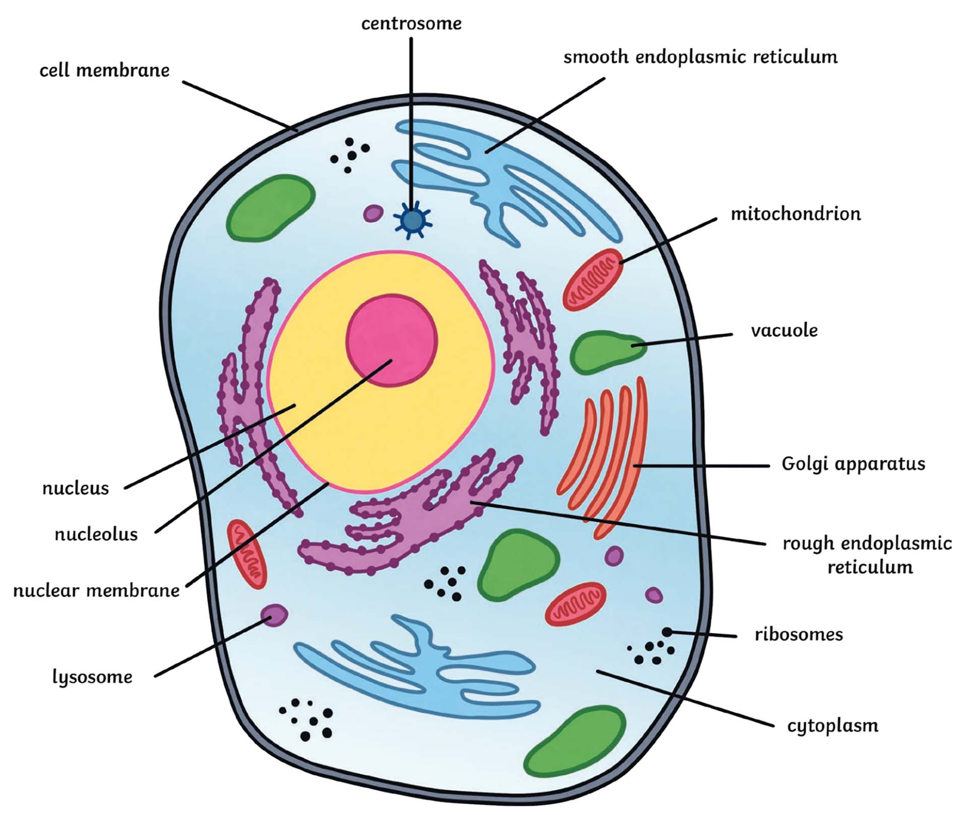
Animal cells and their organelles. Color and abe theVsospmesan. Animal cells are eukaryotic cells that have both a membrane-bound nucleus and other membrane-bound organelles. It makes proteins and lipids that will be exported by the cell.
Smooth ER does NOT have ribosomes on its surface. Both plant and animal cells have double membranes and their own DNA. An organelle think of it as a cells internal organ is a membrane bound structure found within a cell.
Although animal cells can vary considerably depending on their purpose there are some general characteristics that are common to all cells. Color and abe the I orc ryaoñan e Both plant and animal cells have double membranes and their own DNA. Grade 8 Grade 9 Cell parts and functions worksheet pdf.
The Golgi apparatus or Golgi complex is a flattened layered sac-like organelle that looks like a stack of pancakes. Color and label the lysosomes tan. Below you can find a list will all of them animal cell organelles and their functions with and imagediagram to help you visualize where they are and how they look within the cell.
The Golgi body modifies packages proteins and carbohydrates into membrane-bound vesicles for export from the cell. Animal Cell Definition Structure Parts Functions And Diagram The animal cell has 13 different. Plant and animal cells are similar in that they are both eukaryotic and have similar types of organelles.
Nutrients are digested by the cell here as well as old cell organelles that are going to be recycled. Nutrients are digested by the cell here as well as old cell organelles that are going to be recycled. Eukaryotic cells are relatively large cells with a nucleus and specialized structures called organelles.
Prokaryotes are microorganisms that do everything from make cheese to cause strep throat. The most common types of animal cells are. Some organelles that are found in animal cells but not in plant cells are.
Structurally plant and animal cells are very similar because they are both eukaryotic cells. Color and label the Golgi apparatus Golgi bodies red. They both contain membrane-bound organelles such as the nucleus mitochondria endoplasmic reticulum golgi apparatus lysosomes and peroxisomes.
14 rows Cell Organelles. Although both animal and plant kingdom falls under the eukaryotes multi-celled as opposed to prokaryotic which is single-celled animal cells have much more complex structure. Learn vocabulary terms and more with flashcards games and other study tools.
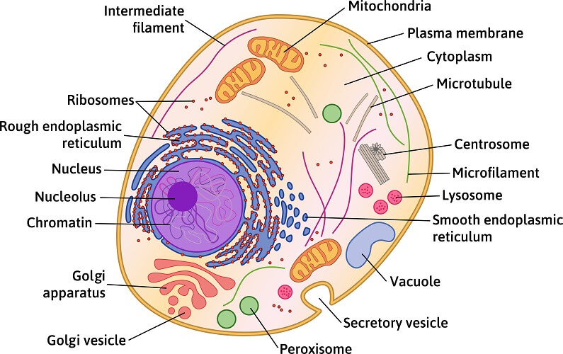
These include structures such as the plasma membrane cytoplasm nucleus mitochondria and ribosomes. A double membrane composed of lipids and. Animal cell organelles and their functions worksheet Learn the names and understand the locations of all the major organelles in an animal cell to have clear concept about its structure.
Two types of cells prokaryotes and eukaryotes. Cells also contain spherical organelles called lysosomes that contain digestive enzymes. Almost all animals and plants are made up of cells.
These organelles carry out specific functions that are needed for the normal functioning of the cell. Unlike the eukaryotic cells of plants and fungi animal cells do not have a cell wall. Animal cells are typical of the eukaryotic cell enclosed by a plasma membrane and containing a membrane-bound nucleus and organelles.
Below the basic structure is shown in the same animal cell on the left viewed with the light. Cells also contain spherical organelles called lysosomes that contain digestive enzymes. This feature was lost in the distant past by the single-celled organisms that gave rise to the kingdom Animalia.
Just like cells have membranes to hold everything in these mini-organs are also bound in a double layer of phospholipids to insulate their little compartments within the larger cells. Click to see full answer. Found in plant and animal cells cell wall ridged outer layer of a plant cell cytoplasm gel.
Start studying Names and Functions of the Organelles in Plant and animal cells.
100+ Animal Cell Diagram Labeled
Start studying Label an Animal Cell. Cellcell wall a cell p label.
Animal Cell The Definitive Guide Biology Dictionary
Posted in Animal Cell Tagged Biology Science Maybe you also like Coloring pages are funny for.
Animal cell diagram labeled. One vital part of an animal cell is the nucleus. A vacuole is an organelle in cells which functions to hold various solutions or materials. Learn vocabulary terms and more with flashcards games and other study tools.
Animal cell diagram labeled vacuole. The cell membrane controls the influx of the nutrients and minerals in and out of the cell. An animal cell diagram is a great way to learn and understand the many functions of an animal cell.
Which of the following organelles does an animal cell NOT have. Image Of An Animal Cell Diagram With Each Organelle Labeled. Cytoskeleton is the.
For instance the roots of the plants help in the absorption of minerals and water. Plant vs animal cells diagram. A worksheet with a simple diagram to label the main subcellular structures Nucleus Mitochondria Ribosomes Cell membrane and Cytoplasm of an Animal cell.
Animal-cell-diagram-not-labeled Tims Printables Mathilda Barnes The significant differences between plant and animal cells are also shown and the diagrams are followed by more in-depth information. What Is An Animal Cell Animal Cell Model Diagram Project Parts Structure Labeled Coloring and Plant Cell Organelles Cake. Printable Animal Cell Diagram Labeled Unlabeled and Blank.
Animal cell diagram detailing the various organelles Though this animal cell diagram is not representative of any one particular type of cell it provides insight into the primary organelles and the intricate internal structure of most animal cells. Well-Labelled Diagram of Animal Cell The cell membrane is a double-layered membrane made up of phospholipids that surrounds the entire cell. It is mainly made up of water and protein material.
Where prokaryotes are just bacteria and archaea eukaryotes are literally everything else. Its the cells brain employing chromosomes to instruct other parts of the cell. Its the cells brain employing chromosomes to instruct other parts of the cell.
The cell membrane is the outer most part of the cell which encloses all the other cell organelles. 5th grade science and biology. Cytosol is the fluid present within a cell that is made up of water and ions such as potassium proteins and small.
A bacteria diagram in actual fact facilitates us to profit more about this single cell organisms that have. Lets go over the individual components of plant cells listed on a diagram such as the one above and. Plant cell picture plant cell labeled plant cell organelles animal cell parts plant cell diagram plant cell project facts about plants plant and animal cells cell wall.
There are two types of cells - Prokaryotic and Eucaryotic. Printable animal cell diagram to help you learn the organelles in an animal cell in preparation for your test or quiz. Plant cells are eukaryotic cells that vary in several fundamental factors from other eukaryotic organisms.
Identify and label figures in Turtle Diarys fun online game Animal Cell Labeling. Plant And Animal Cell Diagram Class 9. A Labeled Diagram of the Animal Cell and its Organelles.
The diagram like the one above will include labels of the major parts of an animal cell including the cell membrane nucleus ribosomes mitochondria vesicles and cytosol. Simple black and white doodle of pedestrians. The cells of animals are the basic structural units for the wide variety of life we see in the animal kingdom.
The mitochondria are the cells powerplants combining chemicals from our food with oxygen to create energy for the cell. The various cell organelles present in an animal cell are clearly marked in the animal cell diagram provided below. Have chloroplasts and use photosynthesis to produce food have cell wall made of cellulose A plant cell has plasmodesmata - microscopic channels which traverse the cell walls of the cells one very large vacuole in the center are rectangular in shape Animal Cells.
Printable animal cell diagram labeled unlabeled and blank. In 5 minutesthis video is specifically for beginnerscontinue f. Eukaryotic cells are larger more complex and have evolved more recently than prokaryotes.
Animal Cell - Science Quiz. Article by Tims Printables. Drag the given words to the correct blanks to complete the labeling.
Animal cell label the parts of the animal cell below. Animal cells are packed with amazingly specialized structures. Animal cells are eukaryotic in nature possessing a nucleus and organelles that carry out the different functions the cell must do to thrive and reproduce.
Plant cell diagram animal cell diagram featured in this printable worksheet are the diagrams of the plant and animal cells with parts labeled vividly. Cells Blank Plant And Animal Cell Diagrams To Label Note Taking Or Assessment Teacherspayteachers Com Plant And Animal Cells Cell Diagram Animal Cell Learn vocabulary terms and more. The cell membrane is the outer most part of the cell which encloses all the other cell organelles.
35+ 7th Grade Animal Cell Model Labeled
3-D Animal Cell Model Project Rubric Grading. Improve your science knowledge with free questions in Animal cell diagrams.

Jacob S 7th Grade Animal Cell Science Project Cells Cute766
On a white piece of paper or poster board draw the outline of your cell and fill.

7th grade animal cell model labeled. A bacteria diagram clearly helps us to profit extra approximately this unmarried cell organisms which have neither membrane-bounded nucleolus or organelles like mitochondria and chloroplasts. One fun way to learn it is by knowing animal cell model ideas. 3-D Cell Model Project 100 points Project Assigned.
This animal cell 3d model helps us to learn about different parts of animal cell. 7th Grade Science - Plant and Animal Cell Vocab. Some of the worksheets for this concept are Label the animal and plant cell organelles and structures Animal cell Cell comparison work Label the human cell Prokaryotic and eukaryotic cells Organelles in eukaryotic cells Ada Cell.
This project will count as a major. 7th grade Science Label Parts of Animal Plant Cell. This is nice way to make 3d model of animal cell school project.
Jelly-like substance found in cells that the. This science project animal cell model with labels helps students to learn about all parts of animal cell in detail. Identify 2 resources for supporting active student learning in science 2.
Colours Poster or Acrylic Paint 6. Displaying top 8 worksheets found for - Label Animal Cells. Plant and Animal Cell Vocabulary.
Plant And Animal Cells 7th Grade Science Plant And Animal Cells Libguides At Amarillo Isd Students can complete the cell diagrams for note taking additional practice or as an assessment. Today we are making animal cell model project for school. - All parts of your cell must be labeled clearly in order to receive credit.
Terms in this set 14 Cell wall. A rigid supporting layer that surrounds the cells of plants and some other organisms. Copy this to my account.
Does not have a cell wall or chloroplast and a small vacuole. Plant Cell And Animal Cell Diagram 8th Standard. If you wish to keep your animal cell project simple you can create a labeled drawing.
By making a 3-D model of the cell the student will become aware of the various organelles and structures which make up a plant or animal cell. Terms in this set 25. O5 Animal cell diagrams.
The structure labeled G give rise to spindle fibers and exclusively seen in animal cell. The cell project is the first project assigned outside of the classroom this year in 7th grade science. The worksheets recommended for students of grade 4 through grade 8 feature labeled animal and plant cell structure charts and cross-section charts cell vocabulary with descriptions and functions and exercises like identify and label the parts of the animal and plant cells color the cell organelles match the part to its description fill in.
Thermocol Styrofoam Sheet 1 inch thick 2. Plant Cell or Animal cell Project Due. Advantages of Working model project for school.
Describe 3 hands-on activities related to cell organelles. Animal cell model labeled. We have made all parts of animal cell and label it using paper flags.
Animal cells do not have cell walls. 16 rows 7th Grade - Cell Parts and Functions. Drawing of an Animal Cell.
From animal cell label worksheets worksheets to animal cell label diagram videos quickly find teacher-reviewed educational resources. As a result of this activity participants will be able to. Label parts and thousands of other science skills.
Feb 7 2019 - ANIMAL CELL MODEL IDEAS Science class is always lots of fun. E-mail to a friend. Learning science can be done in fun ways especially when you learn the animal cell anatomy.
With the cell at center stage guide your seventh grade biologists. An introduction and general requirements for creating a model of a plant or animal cell are provided for basic biology learners.
25 Animal Cell Under Electron Microscope Labelled
Diagram Of Animal Cell Under Electron Microscope Labeled. Label the cell wall cell-surface membrane capsule circular DNA flagella and plasmid.
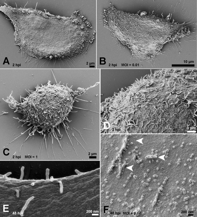
Ultrastructural Analysis Of Sars Cov 2 Interactions With The Host Cell Via High Resolution Scanning Electron Microscopy Scientific Reports
Under the intense radiation of the electron microscope 011 electron per Å 2 the question of viability of cells naturally arises because the amount of radiation absorbed during highmagnification imaging is sufficient to cause cell death.
Animal cell under electron microscope labelled. The cell membrane is important in that. Labeled animal cell under electron microscope 8745961 orig. Animal cells have a basic structure.
Make your work easier by using a label. Describe why it is important to prepare. Endoplasmic Reticulum Rough And Smooth British Society For.
Labelled diagram of a plant cell under microscope posted on march 18 2011 by admin onion cells stained with methylene blue look at the images of onion cells as they would be seen under a microscope draw each magnification label appear high picture plant and animal cell. But at the same time it is interpretive. Labeled Animal Cell Under Electron Microscope.
Draw a labelled diagram of the internal structure of an animal cell as seen with an electron microscope. Animal Plant Cells Gcse Science Biology Get To Know Science Youtube Mitochondrion are visible with a light microscope but cant be seen in detail. Ribosomes are only visible with an electron.
The original and the labelled images are already used world-wide for preparation for exams. Labels are a means of identifying a product or container through a piece of fabric paper metal or plastic film onto which information about them is printed. It is an electron micrograph of cells largest and most important organelle the mitochondria and is characterized by the following features Fig.
Bookfanatic89 Diagram Of Plant Cell Under Electron Microscope. Make your work easier by using a label. Its a thin slice.
Clearly visualized under an electron microscope it must be labeledcell structure the physics teacher april 23rd 2018 - 2 1 cell structure identify the parts of an animal cell as seen under light microscope the existence and definition of prokaryotic and eukaryotic cells me 5 10. So lets begin by drawing a rough-oval shape. See how a generalized structure of an animal cell and plant cell look with labeled diagrams.
Ziehen die pins an die richtige stelle auf dem bild. It is flexible and has pores. However no obvious structural damage.
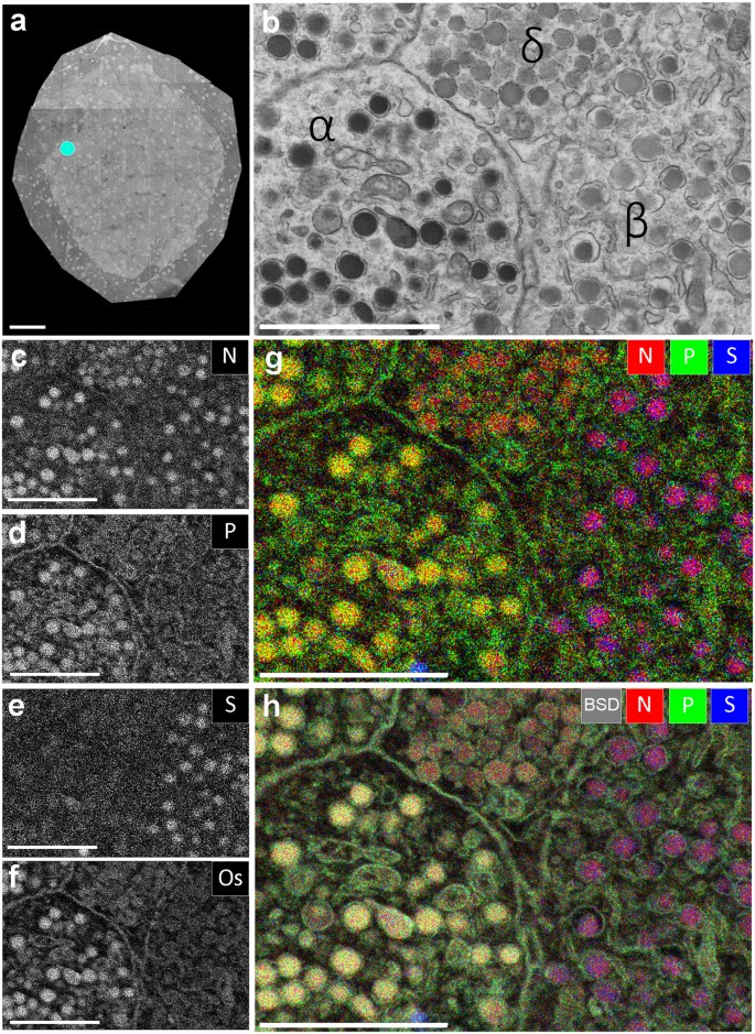
We all do not forget that the human physique is quite problematic and a method I. Diagram Of Animal Cell Under Electron Microscope. Both the global and high-resolution distribution of colloidal gold labels on cells can be readily determined.
Labels are a means of identifying a product or container through a piece of fabric paper metal or plastic film onto which information about them is. The plant cell as more rigid and stiff walls. Posted 6 years ago.
Wide collections of all kinds of labels pictures online. Wide collections of all kinds of labels pictures online. These are both specific types of.
Learn the structure of animal cell and plant cell under light microscope. You see that many features are in common. Here is an electron micrograph of an animal cell with the labels superimposed.
Labelled animal cell diagram gcse. The cell membrane also known as plasma membrane or plasmalemma consists of three layers when viewed under the electron microscope. Electron Micrograph Animal Cell Under Electron Microscope.
A typical animal cell as seen in an electron microscope Medical Images For PowerPoint. Most cells both animal and plant range in size between 1 and 100 micrometers and are thus visible only with the aid of a microscope. Transmission and some scanning electron microscopic images oforgans cells.
Monday April 5th 2021. The three layers are composed of one layer of phospholipid sandwiched between two protein layers. Table D leads to images of electron microscopes or protocols for tissue preparation.
1 The name mitochondria was given by Benda 1898 and their ma n function was brought to light by Kingsbury 1912. Plant Cell Under Electron Microscope Labelled Written By MacPride Tuesday June 18 2019 Add Comment Edit. Draw a labelled diagram of the internal structure of a plant cell as seen with an electron microscope.
Typical Animal Cell Pinocytotic vesicle Lysosome Golgi vesicles Golgi vesicles rough ER endoplasmic reticulum Smooth ER no ribosomes Cell plasma membrane Mitochondrion Golgi apparatus Nucleolus Nucleus Centrioles 2 Each composed of 9 microtubule triplets Microtubules. The information can be in the form of hand-written or printed text or. The diagram is very clear and labeled.
The animal cell is more fluid or elastic or malleable in structure. The lack of a rigid cell wall allowed animals to develop a greater diversity of cell types tissues and organs. Below the basic structure is shown in the same animal cell on the left viewed with the light microscope and on the right with the transmission electron.
2 Each mitochondria in section appears as sausage or cup or bowl shaped structure lined by double. A brief explanation of the. Heres a diagram of a plant cell.
Cell is a tiny structure and functional unit of a living organism containing various parts known as organelles.
71+ Edible Animal Cell Model Cake
Animal cells vacuoles are generally small. Sep 8 2018 - ANIMAL CELL MODEL Making a science job might be uninteresting as well as challenging for some individuals.

Edible Animal Cell Model A Project By Gr 11 Student Youtube
Dec 15 2012 - A cell cake I had to do for Biology class It was a blast to make.

Edible animal cell model cake. 3d Animal Cell Project Edible Cell Project Plant Cell Project Cell Model Project 6th Grade Science Projects Stem Projects School Projects. Vacuoles tend to be large in plant cells. I discovered how fun it was to call fondant Endoplasmic Reticulum and now we ge.
The Incredible Edible Cell STUDENT WORKSHEET Table 1. Cake sprinkles Snakes Peanut MMs. Structure and function.
Watch as I make an animal cell model out of cake and fondantMusic By. The most effective method to have lots of fun while additionally producing a functional scientific research job is by making it. Nov 24 2013 - The kids go to homeschool co-ops once a week and my older daughters group was recently given the assignment to create an edible animal cell.
She chose to make an animal cell cake which is round. The Incredible Edible Cell. We can get plant and animal cell edible projects model of animal cell project ideas and 3d plant cell model cake when we scrolling down our mouse they are best photos related to Cell Cakes with Labels.
Several weeks ago Emily had the opportunity to make a cell cake for extra credit in her science class. More information Animal Cell Cake. Make a cake with icing as cytoplasm different colored icing as cell membrane small pieces of licorice as ribosomes jelly beans as mitochondria whopper cut 12 as lysosome frui Edible Animal Cell Animal Cell Model Diagram Project Parts Structure Labeled Coloring and Plant Cell Organelles Cake.
It had to have all of the cell parts such as the Nucleus Nuclear Membrane Mitochondrion Golgi Bodies etc. Ingredients 1 box vanilla cake mix 1 package mini marshmallows 1 package red licorice 1 container green frosting 2 glazed donuts 1 powdered sugar donut hole 1 Twinkie 1 package orange slices gummy candy 1 package black cookie icing 1 package red licorice. An Edible Animal Cell Model My friends son had an assignment for school where he was required to make a 3d animal cell model.
How to make an edible model of an animal cell. It could be made of any food as long as it was all edible and large enough to feed half of her class. But also for many others scientific research job can truly be entertaining as well as entertaining.
